Musculoskeletal conditions are among the leading causes of disability worldwide, significantly impacting individuals’ quality of life. Among these conditions, joint disorders, such as osteoarthritis, are particularly prevalent, affecting mobility and daily functioning.
What is Knee Osteoarthritis?
Knee osteoarthritis (OA) is a degenerative joint disease that affects around 5.4 million people in the UK1, particularly those over the age of 45. Characterised by the gradual breakdown of cartilage, it leads to pain, stiffness, swelling, and reduced mobility.
Early diagnosis is crucial for managing symptoms and slowing disease progression – many who may be living with knee osteoarthritis have X-rays as part of this diagnostic process.
Recognising the Signs
OA often begins with symptoms such as persistent knee pain, stiffness (especially in the morning), swelling, and a reduced range of motion. Some individuals may also experience a grinding sensation, instability while walking, or visible deformities in advanced stages;some studies have found that up to 40% of patients with knee OA have difficulty walking2.
When diagnosing OA, healthcare providers will enquire about a patient’s symptoms and examine their joints. Although not always necessary, X-rays may be used to rule out other possible causes, such as rheumatoid arthritis or a fractured bone.
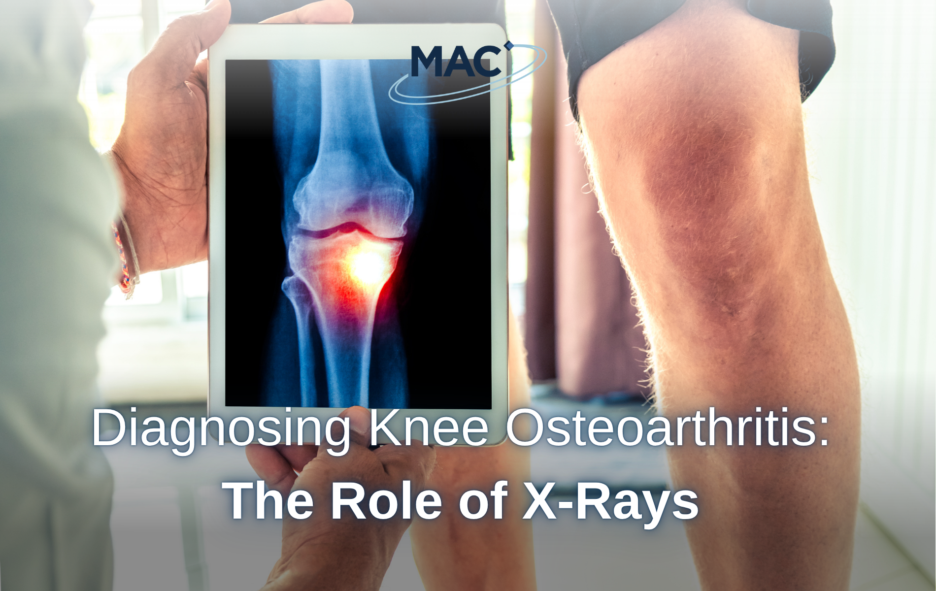
Why X-Rays Are Used
X-rays can provide critical insights into the structural changes within the joint. These include:
- Joint space narrowing: a key indicator of cartilage loss.
- Osteophyte formation: bone spurs that develop around the joint.
- Subchondral sclerosis: increased bone density beneath the cartilage.
- Subchondral cysts: fluid-filled sacs that form in the bone near the joint.
Weight-bearing (i.e., standing) X-rays, particularly the Rosenberg view (taken with the knee bent at 45 degrees), are especially useful. They reveal more pronounced joint space narrowing than non-weight-bearing (i.e., lying down) images, offering a more accurate assessment of disease severity3.
While X-rays may be used in diagnosis, they have limitations as they do not show soft tissues like cartilage, ligaments, or early inflammatory changes.
Improving Quality of Life for Knee OA Patients
MAC Clinical Research understands the challenges that come with living with knee OA and is committed to improving the quality of life for those living with the condition through groundbreaking clinical trials, researching potential new treatments.
Have you had an X-ray to confirm knee OA in the last 4 years? MAC Clinical Research are conducting a new study for a potential OA pain treatment, but we need your help. If eligible you will receive £2010 for your time and commitment, plus reasonable travel expenses will be paid.
If you’re aged between 25 and 79, have moderate or severe OA in your knee, confirmed by X-ray or MRI, and want to take part in a research trial, visit our knee OA webpage to register your interest.
1 Versus Arthritis – The State of Musculoskeletal Health
2 Osteoarthritis and Cartilage – GLA:D® to be walking better: change in self-reported difficulty walking after exercise therapy and education in persons with knee osteoarthritis
3 Radiopaedia – Knee (Rosenberg view)